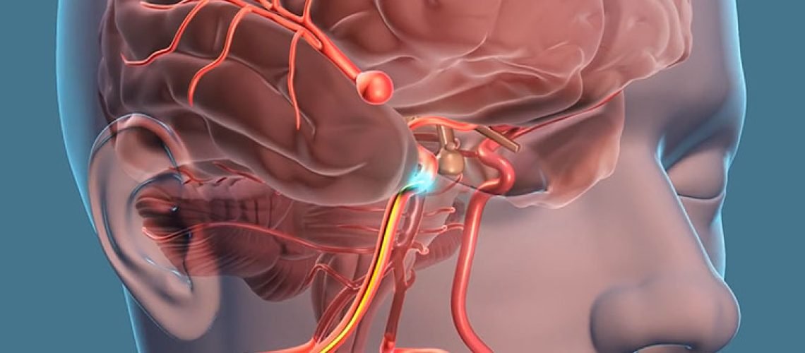
If a CT angiogram or MRI has just told your parent or spouse that they have an aneurysm, take a breath before reading further.
There are several types of aneurysms, but the name on the report matters less than three questions: has it bled, how big is it and where is it, and how healthy is the patient overall. Most aneurysms found on a scan are found without any emergency, and they can be assessed calmly.
Quick answer: An aneurysm is a bulge in the wall of an artery. With no symptoms, the usual next step for an unruptured aneurysm is a planned specialist review of the actual images.
In this guide
- Go to emergency now if you see these signs
- The types of aneurysms, by shape
- Brain aneurysm versus aortic aneurysm
- Which aneurysms are most worrying
- What decides how risky an aneurysm is
- Reading the scan report
- How treatment is chosen
- What to do before the consultation
- Frequently asked questions
- Related guides
- Book a scan review with us
- Sources
As a general typical range, not a fixed rule, that review falls within a few weeks rather than the same day; the neurosurgeon sets the timing for each patient. Red-flag symptoms mean going to an emergency department now (see the emergency section below).
Go to emergency now if you see these signs
Do not wait for an appointment if the person has any of these, whatever the scan said:
- A sudden, explosive headache, often described as the worst of their life
- Repeated vomiting with the headache
- Stiff, painful neck or sensitivity to light
- Fainting, confusion, unusual drowsiness or a seizure
- New weakness on one side, drooping face, trouble speaking, double vision or sudden loss of balance
A ruptured aneurysm causes bleeding around the brain, a type of hemorrhagic stroke, and brain cells begin to die within minutes. Go to the nearest hospital that has CT scanning and neurosurgical support, and take the scan films and reports with you. Call an ambulance rather than driving the patient yourself.
The types of aneurysms, by shape
Shape is the simplest way to tell aneurysms apart. Think of a garden hose.

- Saccular (berry) aneurysm: a small pouch bulging from one side of the vessel, like a balloon on a stalk. This is the most common type in the brain and usually sits where arteries branch at the base of the brain.
- Fusiform aneurysm: the whole width of a segment swells evenly, like a hose widened over a stretch, with no neck or stalk.
- Dissecting aneurysm: the vessel wall layers split and blood tracks between them. This can follow a neck injury or occur spontaneously.
- Mycotic aneurysm: a rare weakening caused by infection of the vessel wall, linked for example to infected heart valves. The word does not mean fungal in most cases.
- Giant aneurysm: not a shape but a size category, generally those larger than 25 mm. They behave differently and need individual planning.
People often ask for “the three types of aneurysms”. There is no single official trio. Usually it means saccular, fusiform and dissecting; others mean location (brain, thoracic aorta, abdominal aorta). We prefer to describe a particular patient by shape, size and place.
Brain aneurysm versus aortic aneurysm
Many pages online mix these up. Most aneurysms in the body are in the aorta, the main artery running through the chest (thoracic) and abdomen (abdominal). Those are managed mainly by vascular and cardiac teams. A brain (intracranial) aneurysm lies in the arteries supplying the brain and is what neurosurgeons assess. If your report says “cerebral”, “intracranial”, “MCA”, “ACA”, “ICA” or “basilar”, it is a brain-artery finding.
Which aneurysms are most worrying
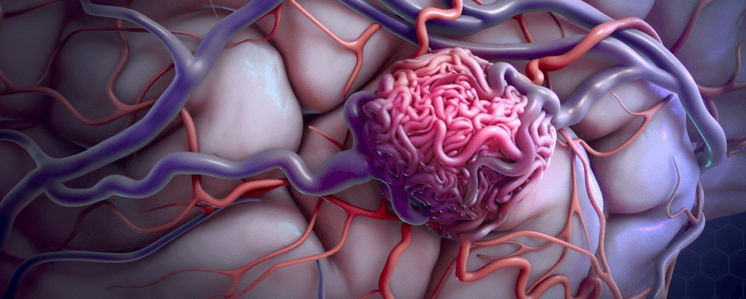
Aneurysms in the neck arteries (carotid or vertebral), which carry blood up to the brain, need specialist assessment even before they bleed, because clots or fragments can travel from them to the brain.
What decides how risky an aneurysm is
We weigh these together, never one alone:
- Rupture status: bleeding changes everything and means urgent treatment.
- Size and shape: larger aneurysms and those with an irregular outline or a small “daughter bulge” generally concern us more.
- Location: some arteries, such as those at the back of the brain, carry higher risk.
- Growth: an aneurysm that enlarges on repeat imaging is treated differently from a stable one.
- The patient: high blood pressure, smoking, family history of aneurysm or bleeding, age and other illnesses all count.
Be wary of anyone quoting a rupture percentage without seeing the scan. Numbers from general studies cannot be applied to one person without the images.
Reading the scan report
A CT angiogram or MR angiogram shows the vessels. Catheter angiography, where a thin tube is guided through an artery, gives the most detailed picture and is sometimes needed to plan treatment. Report words like “saccular outpouching”, “3 mm” or “wide neck” describe shape and size. Words like “subarachnoid hemorrhage” mean bleeding and need urgent action. Online reading cannot replace a specialist review of the images.
How treatment is chosen
| Approach | In brief | Often considered when |
|---|---|---|
| Monitoring | Repeat scans, blood pressure control, stopping smoking | Small, unruptured, low-risk features |
| Endovascular coiling | Catheter from the artery fills the aneurysm with coils | Suitable shape and location |
| Flow diversion | A stent-like device redirects blood flow | Some wide-necked or fusiform aneurysms |
| Surgical clipping | A clip is placed across the aneurysm through an opening in the skull | Anatomy favouring open surgery |
The choice depends on the aneurysm’s size, shape, location, whether it has ruptured, and the patient’s health; the specialist decides case by case.
Not every aneurysm needs an operation, and not every one should be left alone. We would be cautious of anyone who says “surgery tomorrow” or “ignore it” without examining the images.
What to do before the consultation
- Bring the actual films or the CD/online link, not only the printed report, plus earlier scans for comparison.
- List current medicines, especially blood thinners, and any history of high blood pressure, diabetes or heart disease.
- Check blood pressure as advised by the family doctor, and avoid smoking and tobacco.
- Note any family history of aneurysm or brain bleeds.
Questions worth asking: Is it ruptured? What are its type, size and location? Is it stable? Is monitoring reasonable, and how often should we scan? What are the options and risks for this patient? Which warning signs should send us to emergency? Dr. Shyam D’s clinic in Madurai can review the films with you; the Contact Us page at https://drshyam.com/contact-us-2/ lists how to arrange it.
Frequently asked questions
Can an aneurysm be present for years without symptoms?
Yes. Many are found by chance during scans for headaches or other reasons, and cause no symptoms until or unless they press on a nerve or bleed.
Can we travel for a planned review?
A planned trip for a specialist review is reasonable only when the patient has no red-flag symptoms and the scan showed no bleeding. For warning signs at any point, see the emergency section above.
Are aneurysms hereditary?
A family history raises the likelihood, so close relatives of someone with a brain aneurysm should mention it to a doctor. Whether screening is suitable is an individual decision.
Related guides
- When should you see a neurosurgeon? Warning signs you shouldn’t ignore
- Chronic headaches: 7 warning signs you need a neurosurgeon, not just medication
- What does a neurosurgeon do?
Book a scan review with us
If your family member has an aneurysm on a scan and no emergency symptoms, a neurosurgeon can review the actual images with the full history. Dr. Shyam D’s clinic in Madurai offers consultations (see https://drshyam.com/contact-us-2/). Bring the films, the CD or online link, the written report and any earlier scans.
Sources
- Aortic aneurysm — MedlinePlus (U.S. National Library of Medicine); supports the note that most body aneurysms are in the aorta.
- Brain aneurysm — MedlinePlus (U.S. National Library of Medicine); supports the descriptions of brain (intracranial) aneurysms.
- Hemorrhagic stroke — MedlinePlus (U.S. National Library of Medicine); supports the note that a ruptured brain aneurysm causes bleeding around the brain.
This article explains general medical information. It is not a diagnosis — please see a qualified doctor about your own symptoms.